https://www.sciencedirect.com/science/article/abs/pii/S0092867426006410
https://www.cas.cn/cm/202606/t20260622_5112939.shtml
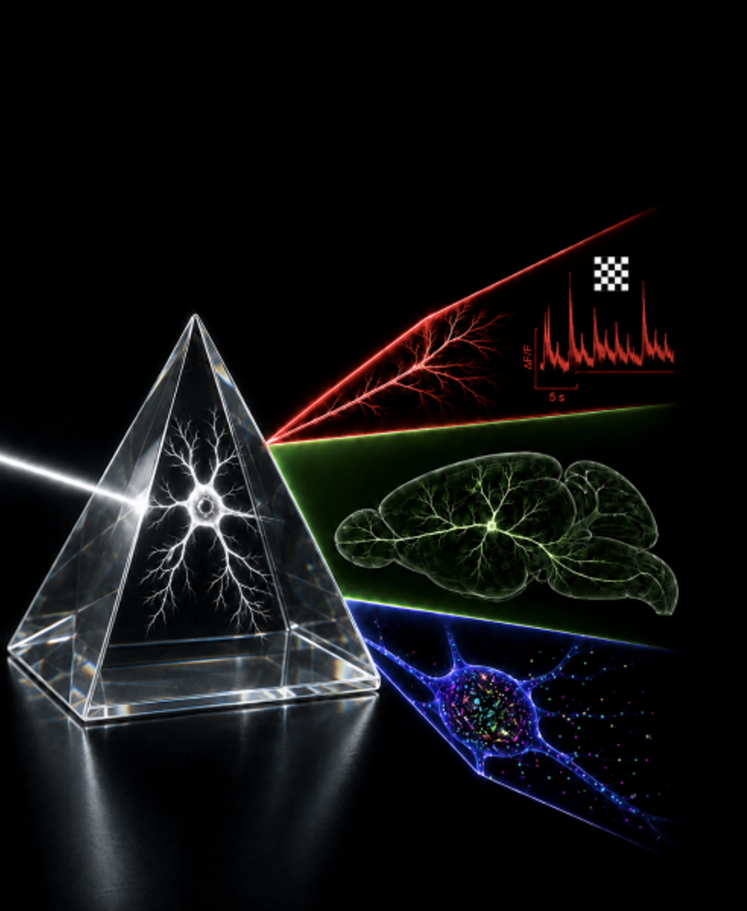
To fully understand the human brain, one must comprehend how neuronal gene expression and morphological connections jointly shape the diverse functions of neurons and construct the brain’s functional neural networks. Recently, a research teams at the CAS Center for Excellence in Brain Science and Intelligence Technology achieved a complete tri-modal analysis of a single neuron.
To understand brain function and tackle brain disorders, it is essential to determine three key aspects of every single neuron:
- function—the dynamic activity of the cell in responding to external stimuli and processing internal information;
- structure—the cell’s morphology and how it connects with other neurons;
- molecules—the gene expression and protein composition within the cell.
The research team developed a novel multi-modal analysis platform named IMC which integrates the entire experimental workflow—from in vivo observation to genetic analysis—enabling the world’s first high-precision, simultaneous analysis of a single neuron’s function, structure, and molecular profile.
The platform relies on two proprietary, patented core technologies:
- A high-resolution, multi-plane, parallelized two-photon microscope capable of fully reconstructing whole-brain neuronal connections without the need to section brain tissue;
- A dual-color expansion fluorescence in situ hybridization (FISH) technique that precisely localizes intracellular gene molecules, allowing for the simultaneous detection of six gene types in a single run.
The experimental process consists of three steps:
- recording in real-time the responses of neurons to visual images and facial movements in awake mice;
- fully reconstructing the neuron’s fiber network as it extends across the entire brain; and
- precisely capturing the distribution and abundance of all genes within the cell. Throughout these three steps, the spatial location of the cell is preserved, enabling precise data alignment and matching.
Using this platform, the researchers have acquired complete tri-modal data for hundreds of neurons, leading to several novel discoveries. Compared to single-dimensional data, integrating cellular morphology with genetic information allows for more accurate prediction of neuronal signal responses. The study also revealed that the intracellular distribution of genes serves as a key marker for distinguishing between different types of neurons.
The research team also identified a previously undefined subtype of excitatory neuron; these cells co-express marker molecules typical of inhibitory neurons and exhibit unique response characteristics to visual stimuli—findings that challenge established academic understanding of neuronal classification.
The multi-modal analysis platform (IMC) enables researchers to comprehensively track a single neuron’s evolution across genetic, morphological, and functional dimensions. It facilitates both the deciphering of the brain’s fundamental computational logic and the investigation of the cellular pathological roots of brain disorders such as Alzheimer’s disease. Furthermore, the homologous 3D data generated by the team can serve as a standard reference database for research in brain science and brain-inspired artificial intelligence. This study fills a critical gap in the experimental tools available to global brain initiatives, providing essential technical support for mapping the brain in its entirety and developing targeted interventions for brain diseases.