https://www.cas.cn/syky/202510/t20251017_5085523.shtml
http://english.cas.cn/newsroom/research_news/life/202510/t20251017_1089587.shtml
https://www.science.org/doi/10.1126/science.ads7954
A team from the University of Science and Technology of China (USTC) and others have used millisecond-level time-resolved cryo-electron microscopy to capture the complete dynamic process of synaptic vesicle release and rapid retrieval. They have proposing a novel “kiss-and-retract/fusion” model as the “microscopic code” for efficient information transmission in the brain.
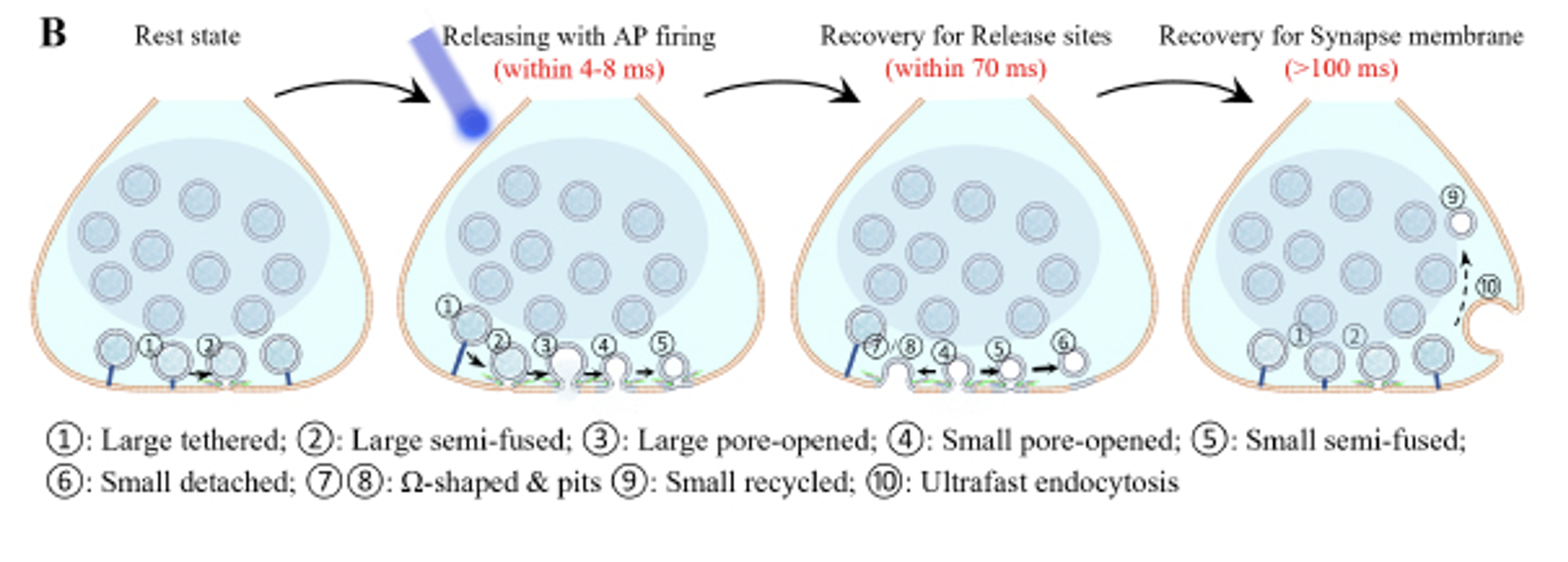
The team expressed a light-sensitive protein in neurons and precisely stimulated action potentials with a laser, triggering synaptic vesicle release. Then, at a set time, the electron microscope grid carrying the sample was rapidly lowered into a cryogen, instantly freezing the cells. By precisely controlling the interval between illumination and freezing, the team released the sample at intervals between 4 and 300 milliseconds, capturing snapshots of the vesicle’s structure at different stages. Based on the analysis of thousands of high-resolution 3D structural data sets, the team discovered that vesicle release and rapid recycling can be divided into a three-stage dynamic process: the vesicle forms a nanoscale fusion pore with the presynaptic membrane (“kissing”), then rapidly shrinks to a smaller vesicle with half the surface area (“shrinkage”). Ultimately, most vesicles are recycled by “escape,” while a few undergo “full fusion.” This intermediate shrinkage is crucial, laying the structural foundation for efficient, high-fidelity signal transmission at synapses.