https://www.cas.cn/cm/202509/t20250918_5082671.shtml
https://doi.org/10.1038/s41586-025-09344-w
In neural interface systems, such as brain-computer interfaces, electrodes serve as the core interface sensors connecting electronic devices and the biological nervous system, forming the heart of the “interface.” However, current implantable electrodes are static, fixed in position and limited to limited data collection. They can be passively attacked by immune responses and even fail to conduct electricity, severely restricting the application and future development of brain-computer interfaces.
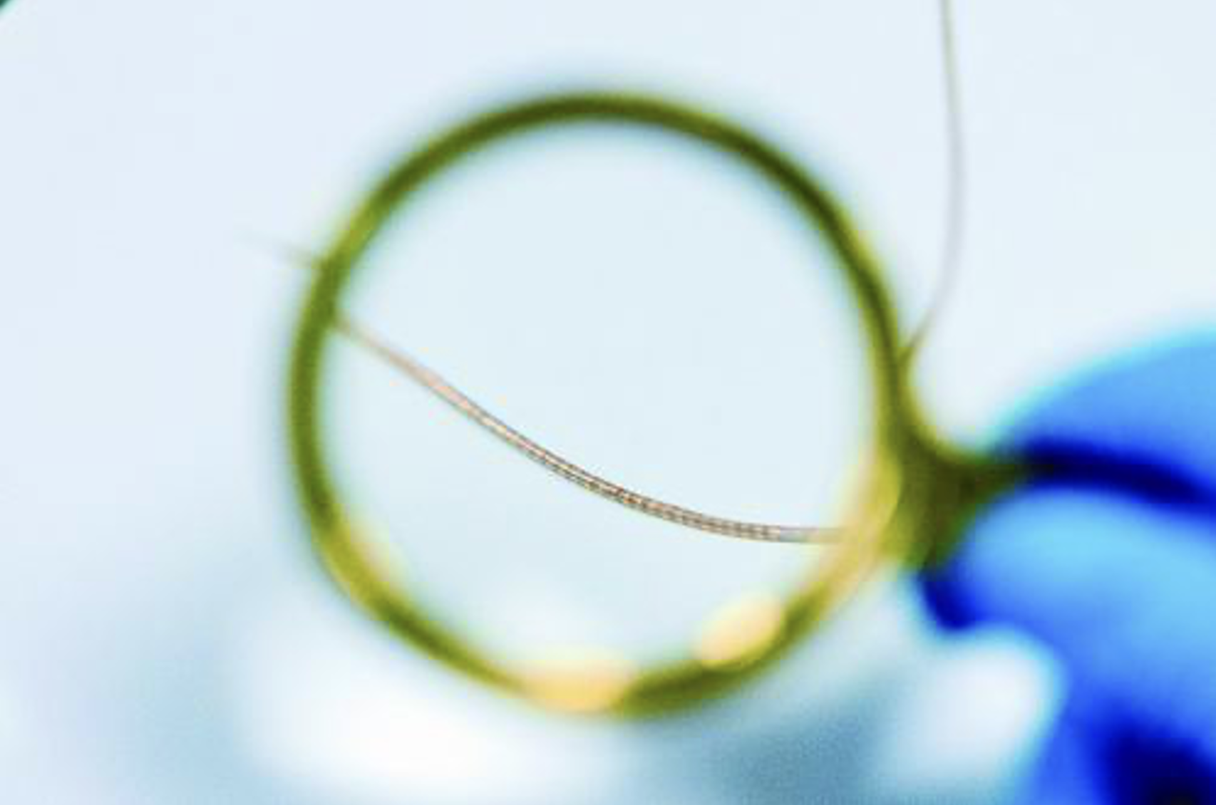
A research team at the CAS Shenzhen Institutes of Advanced Technology (SIAT) has developed a hair-thin, flexible, stretchable, and freely actuable nerve fiber electrode called NeuroWorm. The researchers incorporated tiny magnetic components into flexible electrodes and utilized external magnetic fields to enable them to retain dynamic properties, including adjustability and movement, even after implantation.
A soft, stretchable fiber electrode with up to 60 channels independently distributed along the fiber’s length and a diameter of just 196 microns can move through a tiny magnetic head to one end. Combined with a high-precision magnetic control system and real-time image tracking technology, the electrodes can autonomously control their direction within the body and stably record high-quality bioelectric signals. This “dynamic electrode” was shown to “roam” within a rabbit’s skull, actively changing its monitoring target as needed. The research team named it NeuroWorm.
The NeuroWorm not only opens up new avenues for brain-computer interfaces, but also has applications far beyond the brain—they have achieved the first long-term implantation and stable operation of electrodes within muscle. Compared to the brain, peripheral muscles experience greater deformation and stretching during movement, placing higher demands on the electrodes’ flexibility, durability, and signal stability. NeuroWorm, with its miniaturized and stretchable structure, maintains a close fit within muscle tissue while maintaining high-quality signal acquisition, opening up possibilities for exoskeleton control, rehabilitation assistance, and human-machine collaboration in everyday environments.
Using minimally invasive implantation techniques, the team maintained the NeuroWorm’s stable operation within rat leg muscles for over 43 weeks. Notably, 13 months after electrode implantation, the fiber coating formed around the electrodes averaged less than 23 microns thick, and the cell apoptosis rate in the surrounding tissue was comparable to that of normal tissue, demonstrating excellent long-term biocompatibility. In comparison, under the same conditions, the coating of conventional stainless steel wire electrodes exceeded 451 microns and was accompanied by significant cell apoptosis. Furthermore, controlled by an external magnetic field, the NeuroWorm migrated along the muscle surface, allowing for daily monitoring with alternating positions within a week of implantation.
It is understood that this research is expected to provide new ideas for the fabrication of fiber devices and new tools for brain science research, neural regulation, brain-computer interfaces, and human-computer collaboration. Going forward, the research team will continue to conduct in-depth research in the fields of dynamic flexible electrodes and “active” actively responsive flexible electrodes to advance the development of brain-computer interface technology.